What is the fundus?
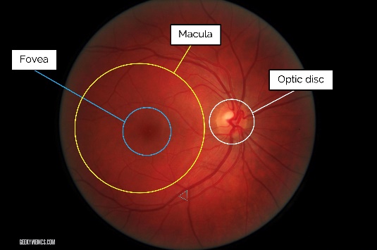
The fundus is a term used to describe the inner surface of the eye. There are differing views on the terminology used to describe the fundus but clinically speaking the main landmarks are the fovea, macula, central retina and peripheral retina. The exact definition of the macula varies, clinicians generally describe it as an area of diameter 1.5mm in the central region.
History of retinal examination
The first written record of something beyond the pupil is Roman author Pliny the Elder’s description of the red reflex, but it was not until much later that deeper exploration into the anatomy behind this was conducted. In the 18th century French surgeon Jean Méry visualised the retinal vasculature of a cat when placed under water. In 1847 Charles Babbage invented an instrument to view the fundus but was discouraged from pursuing this when a distinguished ophthalmologist of the time could not obtain a useful image. A few years later in 1851 Hermann von Helmholtz invented the Augenspiegel (eye mirror), an ophthalmoscope that proved to be the first widespread instrument for examining the internal features of the eye. Over years many innovations have led to the modern techniques described below.
Do I need a fundoscopy?
Dr. Janjua may carry out fundoscopy to screen for eye conditions and diseases that can affect blood vessels. These conditions include:
- Retinal tear or detachment
- Damage to your optic nerve
- macular degeneration- a loss of vision in the centre of your visual field
- Hypertension or high blood pressure
- Cytomegalovirus (CMV) retinitis- infection of the retina
- Glaucoma
- Melanoma - a type of skin cancer that can spread to your eye
What happens during the test?
At the beginning of the procedure, eye drops may or may not be used to dilate your pupils. Next, Dr Janjua will examine the back of your eye. There are three different ways to be examined:
Direct examination: You will be seated in a chair, and the lights in the room will be turned off. Your Dr Janjua will sit across from you and use an ophthalmoscope to examine your eye.
Indirect examination: You will be asked to lie down or sit in a reclined position. Dr Janjua will wear a bright light positioned on their forehead. They will shine the light on your eye while holding a lens in front of your eye to help them examine it.
Slit-lamp examination: You will sit with an instrument in front of you, known as a slit-lamp. It will have a place for you to rest your chin and forehead. Next, Dr. Janjua will turn on a bright light in front of your eye and use a microscope to look at the back of your eye.